
Osteoarthritis of the Knee, Symptoms, Diagnosis and Treatment
Degenerative Joint Disease
- It is the most common of all joint diseases and one of the most common causes of physical impairment.
- It affects both sexes but is more common in women
- It is a disease affecting the cartilage and underlying bone in synovial joints.
- It is characterized by new bone formation (osteophytes) with no load-bearing capacity.
- It is a chronic, progressive disease.
- It is symptomatic in 30% of individuals over 65 years of age.
Risk Factors
- Advanced age
- Female Sexuality,
- Obesity
- Trauma
- Sequence Disorder
- Metabolic, inflammatory diseases
- Connective tissue diseases - Syndromes
Structure of Articular Cartilage
- Cartilage cells (chondrocytes) make up 2-3% of the total cartilage volume, while 65-80% of its content is water.
- Cartilage does not contain blood-lymph vessels and nerve tissue.
- Chondrocytes are the only cell type in articular cartilage and are responsible for the balance between cartilage production and destruction.
Macromolecules in the Structure of Cartilage
- Collagens (95% of its content is type II collagen)
- Proteoglycans
- Non Collagen Matrix Proteins
- Articular cartilage has important functions.
- Reduces the rate of friction.
- It distributes the load on the joint and protects the bone it covers by spreading it over a large area.
- Provides joint stability.
Osteoarthritis (OA) Classification
According to the Number of Joints Involved
- Monoarticular (single joint involvement)
- Oligoarticular (2-5 joint involvement)
- Polyarticular (involvement of more than 5 joints)
According to the Localization of the Involved Joint
- Hip OA
- Knee OA
- El OA
- Spine OA
Symptoms and Signs
- Pain
- Loss of Joint Function
- Joint stiffness
- Joint Swelling and Deformity
- Joint Movement Restriction
- Crepitation
- Muscle Atrophy
Findings
- Sensitivity by touch
- Pain with passive movement
- Crepitation (Sound during joint movements)
- Joint enlargement
- Movement Restriction
- Deformity and dislocation of the joint over time
History
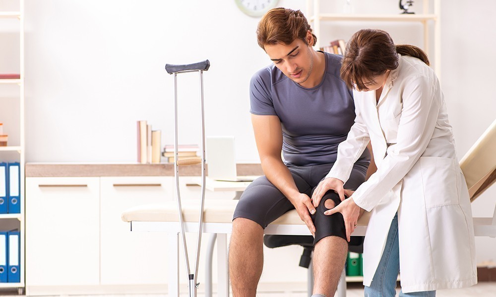
- Physical Examination
- Laboratory Tests
- Imaging Methods
Imaging Methods
X-ray: Although plain radiographs are not very sensitive, they are easier to obtain and relatively inexpensive, making them the most useful imaging modality in the diagnosis of O.A.
Ultrasonography: Its advantage is that it is cheap, easy to use and has no radiation-induced side effects.
In the identification of osteoarthritis, it detects an increase in joint fluid and thickening of the tissue that produces the joint fluid (synovium), especially in hard-to-reach areas such as the hip.
MRI and Computed Tomography: It is a detailed examination and is used to visualize the stage of cartilage damage, subchondral edema, cyst, osteonecrosis.
Radiological Findings
Osteophytes (new bone formation) : These are new bone formations that often occur adjacent to unhealthy cartilage in areas exposed to stress.
Joint space narrowing: It often occurs in load-bearing joints.
Bone Remodeling: It is the response of the bone to the increased load due to the deterioration of the load absorption properties of the cartilage in the damaged area.
Bone Cysts : They are not true cysts. They range in size from a few mm to a few cm with sclerotic, oval and rounded edges.
Free Body in the Joint(Loose Body): The Loose Body occurs as a result of the bone and the cartilage part on it separating from the articular surface and falling into the joint cavity and causes local inflammatory reaction and mechanical findings (instability and stiffness).
Deformity and Partial Dislocation of the Joint:
- It is seen in advanced stages of osteoarthritis.
- Angular deformities develop in osteoarthritic joints, especially in the load-bearing joint.
- Inward bending is most common.
Treatment
- Eliminating causative factors
- To relieve symptoms and comfort the patient
- Making the patient functional
- Protecting the area from trauma and overloads
- Protecting other weight-bearing joints
- Treating the diseased joint and, if necessary, the upper and lower joints with surgical or non-surgical methods
Non-Surgical Treatment Methods
- Cold application
- Use of anti-inflammatory drugs
- Antidepressant drugs
- Anti-inflammatory cytokines
- MMP inhibitors
- Strengthening and range of motion exercises
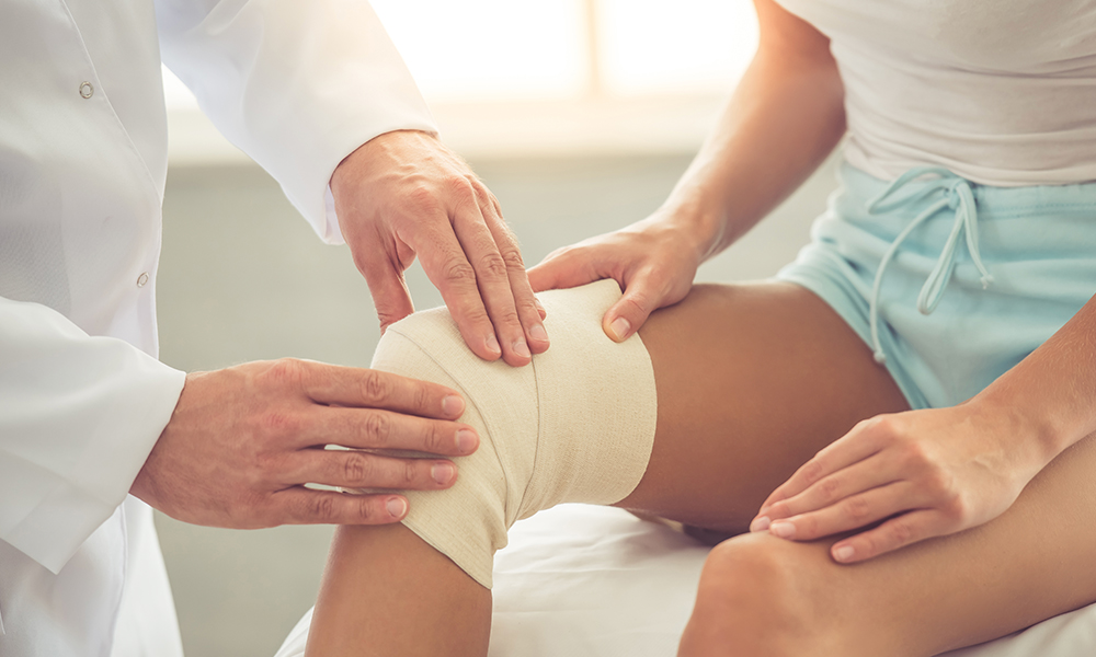
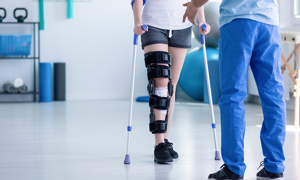
- Stabilization of the joint (splint, collar, brace, orthoses, rest splint, cast, knee brace, elbow brace, ankle brace...)
- Heel Wedge
Intra-articular Treatments
- Stem Cell Applications
- Viscosupplementation (hyaluronic acid, peptide injections)
- Corticosteroid
- Platelet-rich plasma (PRP)
Surgical Treatment
- Arthroscopic (Closed Surgery)
- Partial Knee Replacement
- Total Knee Replacement
- Resurfacing
- Bone surgery to realign the joint